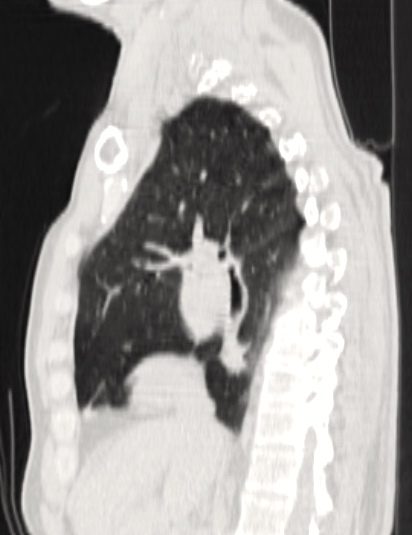
This video presents the case of a 71-year-old male patient, who was diagnosed with a centrally located left lung tumor. Endobronchial ultrasound-guided needle aspiration confirmed an adenocarcinoma. PET-CT revealed no lymph node or distant metastasis. After the chest CT, the authors determined that a pneumonectomy was the only option for surgical resection (Figure 1). Spirometry was suboptimal (FEV1: 58%), but VO2-max was normal (20.4 mL/kg/min). A single incision VATS (SIVATS) approach was used for a left pneumonectomy and lymph node dissection in this patient.
 |
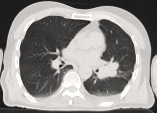 |
| Figure 1A: Lateral CT scan. | Figure 1B: CT scan. |
The length of the incision (7 cm) used in this approach is comparable to that of a muscle-sparing lateral thoracotomy incision. However, the postoperative course is associated with less pain and earlier patient mobility, compared with a thoracotomy approach. Avoidance of two additional intercostal incisions, and potential injury or postoperative pain due to instruments inserted through the intercostal ports, in the SIVATS approach could make this preferable over standard 3-port VATS for pneumonectomy.

Nice work, how do u keep the port stable in the lower part of incision?
We use trocar only as a passage in inserting the camera inside the chest. Then, it is slipped back over the shaft of the camera leaving the incision to other instruments maneuvering freely.